Singing Hatsune Miku

BIOLOGY FORM 5

Powered by Blogger.
RSS

PRESENTATION

  • Digg
  • Del.icio.us
  • StumbleUpon
  • Reddit
  • RSS

FUN EXPERIMENT!!! -LAVA LIGHT

WE ARE... PANTHERA TIGRIS!

WITH OUR AMAZING BIOLOGY TEACHER!!! 


Firstly, you need these...
A) EMPTY BOTTLE
B) VEGETABLES OIL
C) FOOD COLOURING
D) GLITTERS
E) TORCH LIGHT
F) ALKA-SELTZER TABLET


1. Ruth is adding the distilled water in the bottle


PLEASE WATCH OUR VIDEOS TO SEE MORE ABOUT THIS EXPERIMENT... THANKS!! :)


PART 1

PART 2


  • Digg
  • Del.icio.us
  • StumbleUpon
  • Reddit
  • RSS

EXPERIMENT-COW HEART DISSECTION


Cow Heart Dissection Walk Through

The heart dissection is probably one of the most difficult dissections you will do. Part of the reason it is so difficult to learn is that the heart is not perfectly symmetrical, but it is so close that it becomes difficult to discern which side you are looking at (dorsel, ventral, left or right). Finding the vessels is directly related to being able to orient the heart correctly and figuring   out which side you are looking at.The heart is also difficult because the fatty tissue that surrounds the heart can obscure the openings to the vessels. This means that you really must experience the heart with your hands and feel your way to find the openings. Many people will be squeamish about this, and because the heart is slippery, it is easy to drop. Don't be shy with the heart, use your fingers to feel your way through the dissection.

student holding heart

1. Step One: Orientation

When you first remove your heart from the bag, you will see a lot of fatty tissue surrounding it. It is usually a waste of time to try to remove this tissue. Grab some colored pencils to help you identify and mark the vessels you findThere are a few clues to help you figure out the left and the right side, but often the packaging and preserving process can cause the heart to be misshapen. If you are lucky, the heart will be nicely preserved and you will see that the front (ventral) side of the heart has a couple of key features: 
1) a large pulmonary trunk that extends off the top of it 
2) the flaps of the auricles covering the top of the atria. 
3) the curve of the entire front side, whereas the backside is much flatter. The first image shows the front side of the heart, often identified by the coronary sinus that runs cross it at an angle (yellow).

heart

The auricle is the flap that covers the atrium, it looks like an ear. The pulmonary trunk is the located at the front of the heart and enters at an angle.

heart

Step 2: Locate the Aorta

Use your fingers to probe around the top of the heart. Four major vessels can be found entering the heart: the pulmonary trunk, aorta, superior vena cava, and the pulmonary vein. Remember that if you are looking at the back of the heart, then the right and left sides are the same as your right and left hand. This picture was on the board the day of the dissection so that you could glance up and recall which vessel entered which part of the heart.

heart drawing

If you find the pulmonary vein, the aorta should be situated a little bit behind it. It may be covered by fat, so use your fingers to poke around until you find the opening. Push your finger all the way in and you will feel inside of the left ventricle. The left ventricle has a very thick wall, unlike the right ventricle. Insert your finger through the pulmonary vessel to feel the left ventricle and you will notice and feel that it is much thinner than the left side of the heart.
With your fingers or probes in the aorta and the pulmonary trunk you should notice that they criss-cross each other, with the pulmonary trunk in the front.

vessels

At this point, you may want to use your colored pencils to mark these vessels so that you don't get them confused when you are searching for the other two openings that top of the heart.

Step 3: Locate the Veins

The two major veins that enter the heart can be found on the backside, as both enter the atria. On the left side, you should be able to find the opening of the pulmonary vein as it enters the left atrium. The superior vena cava enters the right atrium. In many preserved hearts, the heart was cut at these points, so you won't see the vessels themselves, you will just find the openings. Again, use your fingers to feel around the heart to find the openings. If you've marked the aorta and pulmonary then you won't mistake them for the veins you are looking for. This picture shows all of the vessels labeled.

vessels

Sometimes, the aorta still has its branches attached to it. There are three vessels that branch from the aorta: the brachiocephalic, left common carotid and the left subclavian. The majority of the time, these vessels are not visible because the aorta was cut too close to the main part of the heart when the heart was removed from the animal. Occassionally, you can find the brachiocephalic artery attached, as it is in this photo.

pulmonary trunk

Step 4: Make the Incisions

Now that you have all of the vessels located and marked, you can now open the heart to view the inner chambers. Use the superior vena cava and pulmonary vein as guides for where to cut. You are basically going to be cutting each side of the heart so that you can look inside. (Some dissections will ask you to make a coronal cut where a single cut opens the entire back side of the heart). The heart below is marked to show you where the two incisions should be made.

  heart cut 1

 heart cut 2

Optionally, you may cut the heart in half to expose the chambers. My friends affectionally call these two variations the "hot dog cut" as pictured above because it looks like a hot dog bun, or the "hamburger cut" like that pictured below.

Step 5: Viewing the Chambers

At this point it is helpful to have two hands, one to hold the heart apart so you can take a peak inside of it and another to use a probe to locate the specific parts. Your colored pencils you used to mark the heart in step 2 can also now be used to see where those vessels connect within the heart. For instance, the aorta pencil can now be seen ending in the left ventricle. You can also now see how much thicker the walls of the left ventricle are compared to the right ventricle.

chambers labeled

The other obvious structures seen within the heart are the chordae tendinae which are attached to papillary muscles. These tendons hold the heart valves in place, sometimes they are called the "heartstrings". The valves were probably cut when the heart was opened, but if you follow the "cords" they should lead you to a thin flap that is the atrioventricular 
(bicuspid) valve. You can find a similar valve on the right side of the heart (tricuspid).

heart bicuspid

                   Close-up of valve with chordae tendinae

heart valves


COW HEART DISSECTION (VIDEO)


  • Digg
  • Del.icio.us
  • StumbleUpon
  • Reddit
  • RSS

EXPERIMENT-LAMB HEART DISSECTION


BIOLOGY EXPERIMENT
Lamb Heart
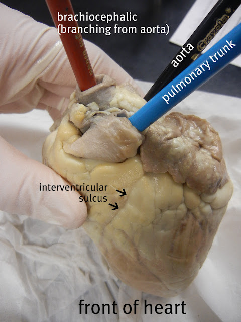
The heart is an oblong muscular organ, which is attached to the lungs and to the two major blood vessels of the body (the aorta and vena cava again) by a tangle of tubes at the top. There are also two “flaps” lying alongside the tube tangle which bear a vague resemblance to the external flaps of the ears, and which share their name: auricles. The probe in the following two pictures indicates the auricles.
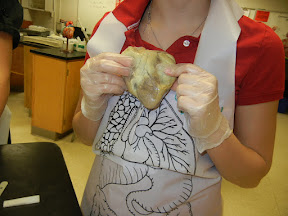
A Lamb Heart
A Lamb Heart

The Tube Tangle
The Tube Tangle

If you wish to slice through a heart to expose the interior, which way should you slice? If the auricles indicate the “sides” of the heart, then a good choice for which way to slice is sideways, through each auricle, so you can see what is inside underneath each auricle. In the heart shown below, I sliced through one auricle, but missed the other. The second auricle is hidden behind the top of the heart on the right hand side.

The Interior of a Lamb Heart

The heart is hollow inside, with two chambers, one underneath each auricle. These are the ventricles. The larger of the two, with thicker more powerful walls, is normally on the left side of the body and is called the left ventricle, and the smaller of the two, with thinner walls, is the right ventricle. (If the heart is made to work by the muscular walls, which of the two chambers do you suppose has the harder job to do?) Within and underneath the auricles are two floppy chambers that look like either the upper portion of the ventricles, or maybe separate chambers atop each ventricle. These are the atria. (The right atrium is hidden in the picture above.) A little poking and lifting with a blunt probe (or an old pencil or pen) can reveal tough thin fabric flaps separating the ventricles from the atria. (When these flaps are lying flat against the walls, each atria and ventricle looks like a single chamber.) The visible tendon-like strings apparently hold down the edges of the flap for some reason. If you gently insert the probe through the various tubes in the top of the heart, it will emerge within one of the chambers. All of the tubes on top of the heart open into one of the chambers within.
(The flaps between the atria and ventricles are the mitral valve and the tricuspid valve–one-way valves that let blood flow from the atria to the ventricles, but not vice versa. The tendons–the chordae tendinae, or heart strings–are for holding the valve steady when it is closed so that it doesn’t blow inside out like an umbrella in the wind. The little lumpy muscles lining the interior walls that pull on these tendons are the papillary muscles.)

DISSECTION OF CHICKEN HEART (VIDEO)


  • Digg
  • Del.icio.us
  • StumbleUpon
  • Reddit
  • RSS

GROUP MEMBERS INFO

Name : Nur Sabrina binti Samsudin
Age : 17 years old
Birth date : 13 July 1998
School : SMK Infant Jesus Convent, Melaka.
Hobby : Shopping
Favourite food : Spagetti
Favourite beverage : Ribena

Name : Jasmine Ho Chia Shin
Age : 17 years old
Birth date : 21 January 1998
School : SMK Infant Jesus Convent, Melaka.
Hobby : Cooking
Favourite food : Western Food
Favourite beverage : Milo Ice

Name : Ruth A/P S. Daniel
Age : 17 years old
Birth date : 24 February 1998
School : SMK Infant Jesus Convent, Melaka.
Hobby : Reading
Favourite food : Fried Chicken
FAvourite beverage : Coke

Name : Ho Jun Er
Age : 17 years old
Birth Date : 15 April 1998
School : SMK Infant Jesus Convent, Melaka.
Hobby : Sleeping
Favourite food : Japanese Food
Favourite beverage : Fruit Juice

  • Digg
  • Del.icio.us
  • StumbleUpon
  • Reddit
  • RSS